SICAT Implant - Getting started. Import of DICOM Data & General Navigation
- Description
- Curriculum
- FAQ
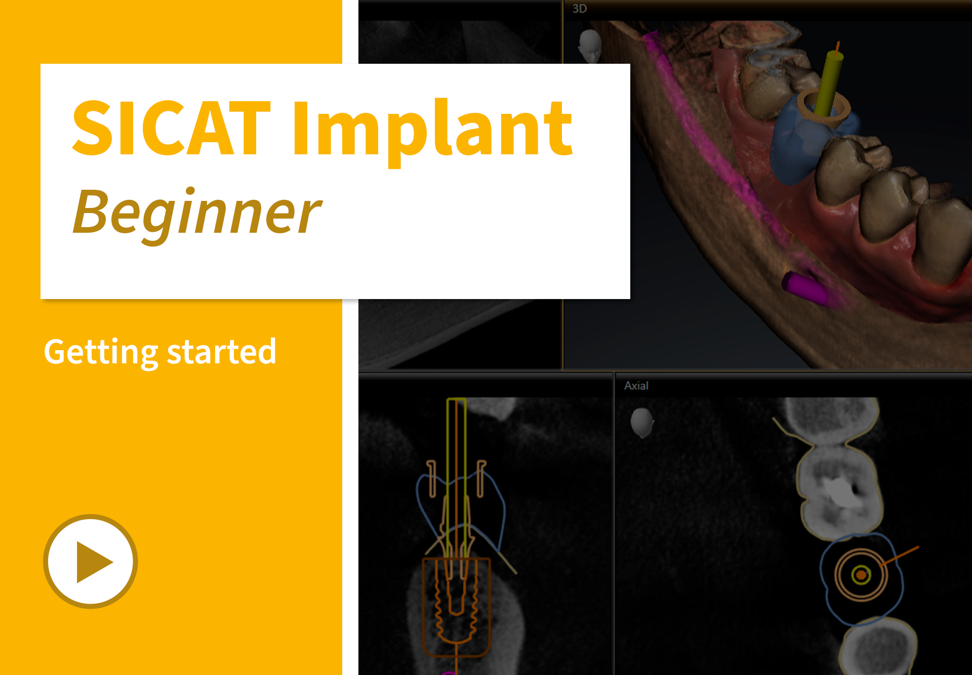
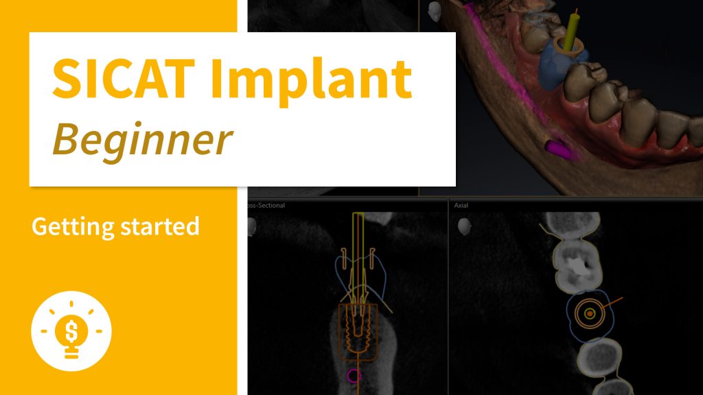
Learn in this course how to use the basic functions of the intuitive software workflow.
The course aims to provide you with a quick introduction to the proven user interface.
In addition to importing DICOM data and adjusting gray values, we will show you how to optimize the visualization in the software individually for your purpose. Use existing CBCT data without the need to convert these.
Finally, learn how to navigate through the software and how to optimally use the different views.
After this course, you will be able to navigate the software self-sufficiently and you are ready for future 3D implant planning cases.
Contents
- How to import DICOM Data
- How to adjust gray values
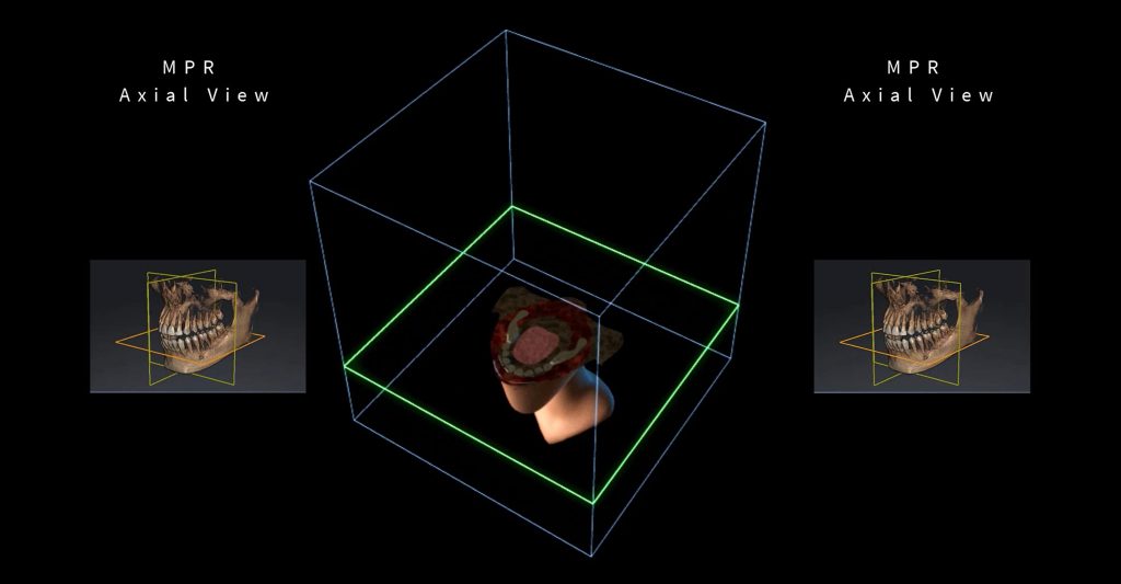
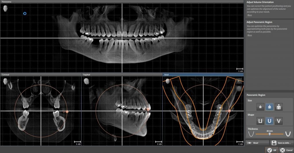
- How to optimize the visualization
- How to navigate through the software – Part I
- How to navigate through the software – Part II
- How to customize SICAT Implant
Target Group
Dentists with no or less experience in using a 3D implant planning software in general and dentists who are not yet familiar with the SICAT Implant software. However, this course is interesting for everyone, who simply wants to learn more about SICAT Implant.
Additional information
Last Update: 2022-08-31
-
1Importing DICOM data
How to perform implant planning based on optical impressions.
-
2Adjusting gray values
How to achieve optimum viewing quality.
-
3Optimizing visualization
How to adjust patient orientation and panoramic curve.
-
4Navigating within the software - Part 1
How to use different views available.
-
5Navigating within the software - Part 2
How to slide through scans and visualize areas for inspection.
-
6Customizing SICAT Implant
How to configure the software to fit your individual needs.


